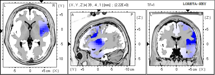
LORETA can be done in a few ways… as a source analysis for the entire EEG or segment being analyzed… or after a decomposition into the ICA components… The later will provide a better estimate of generators of the
individual components which are all blended for the overall LORETA, distorting the sources with other sources.
LORETA
Medication Failure: EEG/qEEG Findings Provide Evidence
This is the PowerPoint “Medication Failure: EEG/qEEG Findings Provide Evidence” as presented at ISNR Conference Workshop 20 September 21, 2013
Presented by:
Ronald J. Swatzyna, Ph.D., L.C.S.W.
The Tarnow Center for Self-Management
drron@tarnowcenter.com
Vijayan K. Pillai, Ph.D.
The University of Texas at Arlington
pillai@uta.edu
Current Research Regarding Blast Injuries in Veterans

This current research from the New England Journal of Medicine – Detection of Blast-Related Traumatic Brain Injury in U.S. Military Personnel – shows that Blast Injury is not at all like mild traumatic brain injury, since the mTBI does not involve white matter injuries. The research does show white matter changes during the medical evacuation, done in Germany using Diffusion Tensor Imaging, and also that the white matter changes continue to evolve. They also show that not all symptomatic blast injuries are seen with this technique.
No traditional structural neuroimaging was able to see this damage (like CT or routine MRI). The NY Times recently reported on soldiers injuries evading the M.R.I and CT Scans
The brain areas involved included the orbital surfaces of the frontal lobe and the temporal areas.
These results point to the need for a clinical diagnosis, not a reliance on any given technology to answer the clinical question.
The endocrine changes from supposed pituitary injury, and the presence of micro-emboli due to pressure wave impact on the thorax that are reported in blast injury is not at all dismissible with these findings.
International Society for Neurofeedback & Research (ISNR) 18th Annual Conference
International Society for Neurofeedback & Research (ISNR) 18th Annual Conference
Denver, Colorado Sept 30-Oct 3, 2010
![]()
ISNR invites you to their 18th Annual Conference for Health Professionals, Education Professionals, Researchers & Students. This conference offers workshops by the leading clinicians and researchers in the field of neuroscience. There will be many workshops and keynote talks on clinical as well as theoretical applications in the neuroscience field.
Derived Feedback Metrics such as Z-score Training
As the technologies advance and the software speed starts to allow derived measures to be used for feedback, the field is being offered many new tools for neurofeedback, including ICA based feedback, LORETA based feedback, and Z-score feedback.
All of these new tools will require clinical validation prior to being able to be considered standard techniques within our field’s armamentarium of efficacious techniques and clinical applications. All of these techniques offer great hope at this time with preliminary results, but careful clinical outcome studies remain to be performed.
In this brief note I will discuss Z-score feedback. This promising technique offers to set normative boundaries around the mean of many features of the EEG, and allow feedback to be controlled by these parameters. This obviously offers great hope to clinical outliers, as their Z-score divergence should be related to their pathology. One difficulty is that database Z-scores also show divergence when an adaptive or counter-balancing feature is used to cope with an abnormal finding. A crutch is not a normal finding, but you can’t walk without it if you have a broken leg.
Thalamic Involvement in the Generation of the Alpha Rhythms
Alpha… it’s not a simple idling rhythm… let’s look at alpha generators:
The thalamic involvement in the generation of the alpha rhythm is being under-valued when looking at the LORETA images of alpha current source generators. The alpha power may come from the sources that LORETA identifies, but the thalamus is intimately involved in alpha rhythm generation, and this is not part of the LORETA image of the sources.
The polarization within the thalamus sets the base frequency of the alpha, but the cortical rhythm requires a complex multi-layer feedback loop from the thalamus to the cortex, and back to the thalamus. Without the cortex, there is a total disruption of the normal spatio-temporal distribution of the alpha wave’s spike trains within the thalamus, and cortical damage often disturbs coherence due to this mechanism.
The thalamus distributes the alpha posteriorly via specific sensory relays, which have a simple return circuit. Like the white matter relay from the lateral geniculate of the thalamus to the occipital lobe’s primary visual areas, and directly back. This thalamo-cortical-thalamic loop is relatively faster than the loop seen frontally. The frontal return circuitry is not simple, but the descending routes are complex and somewhat circuitous, taking more time, and thus it is common for the frontal lobe’s alpha to be at the slower end of the individual’s alpha frequency range. The frontal lobe has a return path through the striatum.
Dementia and Alzheimer’s Disease: LORETA findings
Thanks to Jay Gunkelman who made a very informative post on January 27 on this forum entitled Dementia and Alzheimer’s Disease. There he described the EEG patterns that we should expect and detect when evaluating for AD or other dementias.
I’d like to just throw out there a few other findings that were discovered in a few exploratory investigations while working on some studies with our colleague Alicia Townsend, at the time at Univ. of North Texas. Lexicor funded these projects and now the arrangements are such that I can’t disclose more than was published in the abstracts from our talks at ISNR and AAPB. I did at least want to point to these very preliminary findings because theoretically they are in concert with your explanations.
First, we explored 10 participants between the ages of 65 and 85 were recruited at the University of North Texas Health Science Center. Each was diagnosed by the Alzheimer’s Disease Assessment Scale and a medical interview. The aim of the study was to identify current source density markers in AD. EEG recording of the eyes closed condition of an AD group was compared to an age-sex matched control group using within-subject multiple t-test procedures. sLORETA difference maps in nine frequency bands were investigated. Interestingly the results showed that there was a significant increase in current source density in the delta and theta bands in the Brodmann Area (BA) 39 of the right temporal lobe and BA 31, the cingulate gyrus respectively. Additionally there were decreases in alpha in the BA 21 of the right temporal lobe and right inferior parietal lobule (Sherlin, Townsend & Hall, 2006).
A discussion on LORETA software use and licensing.
April 30, 2009
Leslie Sherlin, PhD
There recently has been some discussion regarding the use of low resolution brain electromagnetic tomography or LORETA, sLORETA and eLORETA. I felt compelled to make a few comments regarding this since there may be some confusion of how LORETA works and the usage of LORETA as an inverse solution specifically the licensing agreements of the KEY Institute for Brain-Mind Research at the University Hospital of Psychiatry, Zurich.
My intention is to very briefly explain the license agreement so that the end user can be informed. I’ll do so in an informal way by telling the story of the implementation of these methods from my perspective. For a more formal description of the use of LORETA families and some examples you can see a recently written chapter 4 by myself (Sherlin, 2009) in the latest edition of the book Introduction to Quantitative EEG and Neurofeedback edited Budzynski, Budzynski, Evans & Arbarbanel.
In 2000 I had the great privilege to visit with Roberto Pascual-Marqui PhD, the developer of the LORETA family, with my colleague and fellow student Marco Congedo. At this time the LORETA-Key software (Pascual-Marqui, 1994, 1999), had not been widely distributed and utilized in the United States. Marco had significant interest in using LORETA for visualizing brain activity and for exploring newer methods for neurofeedback and had many questions for Roberto. So upon the invitation of Roberto, Marco found funding to travel to Zurich and learn the details from the creator and I happen to be standing in the right spot at the right time. Roberto Pascual-Marqui trained us extensively on how to use his software, named LORETA-Key, which had been already released as free academic software. The LORETA-Key software is a collection of independent modules that the user must run in sequence in order to get from raw EEG to LORETA images.

