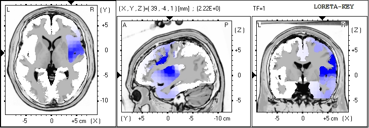
LORETA can be done in a few ways… as a source analysis for the entire EEG or segment being analyzed… or after a decomposition into the ICA components… The later will provide a better estimate of generators of the
individual components which are all blended for the overall LORETA, distorting the sources with other sources.
 Jay Gunkelman
Prof. Juri D. Kropotov Interview 2013
Juri speaks about the recent ERP meeting in St Petersburg Russia, held during the summer’s “white nights” when the night sky does not darken fully and the night is very short. Juri is interviewed in his offices, only 200 meters from Pavlov’s famous laboratory.
The discussion of the new methodology of ICA decomposition of the ERP, as well as the benefit of ERP added to the EEG/qEEG is discussed. There is discussion of the diagnostic specificity of the ERP methods, and the concept of biomarkers.
National Institute for Health and Clinical Excellence (NHS NICE) has published final guidance recommending the use of brain monitoring technology
The healthcare guidance body NICE (National Institute for Health and Clinical Excellence (NHS NICE) has published final guidance recommending the use of brain monitoring technology such as the Bispectral Index (BIS, Covidien), E-Entropy (GE Healthcare) and Narcotrend-Compact M (MT MonitorTechnik GmbH & Co). These EEG-based depth of anaesthesia monitors should be considered as positive options in patients receiving total intravenous anaesthesia (TIVA) and in patients who are considered at higher risk of adverse outcomes during any type of general anaesthesia, such as seniors, those with high body mass index, and those with cardiovascular and liver disease.
EEG based Brain Monitoring Systems helps clinicians assess patient consciousness levels through measuring the electrical activity in the brain. This includes patients who are at higher risk of unintended awareness (anesthesia too light) and also those patients who are at higher risk from excessively deep anaesthesia.
Superior surgical outcomes, the low cost of the testing and the ease of use of these technologies all contribute to the recommendations.
You can access these documents through ASET (www.ASET.org)
Move up to modern de-artifacting
There is an on-going dispute regarding de-artifacting methods used in qEEG. Though there are vested interests counseling against the use of modern techniques to remove artifact while leaving the underlying EEG intact, there are also those who have specialized in the area that can provide a detailed reply to the vested interests. Just such a reply was posted recently in a commercial list server, and we got the author’s permission to re-post the discussion on the qEEGSupport.com website in a non-commercial publicly accessible form for all to see.
It specifically points to the fact that the phase changes seen are due to removal of artifact, not the distortion of the underlying EEG, which has residual subtle artifacts remaining if processed with classical approaches.
If you cut time segments out of the EEG to remove artifacts, you also remove the underlying connectivity information, splicing discontinuous microstates together destroys the underlying time series.
In the give and take of the real world of neuroscience, the need to provide a valid time-series showing the connectivity of the neural networks, yet free of artifact, is driving the need to switch to more modern techniques than snipping out segments of time. If you want to distort the timeline of the EEG (phase) just cut and paste lots of EEG together in one second chunks.
The neuroscience community will undoubtedly continue to discuss these issues, but the need for clean valid EEG is driving the field to these newer techniques, and they are performing well under the scrutiny.
Jay Gunkelman
Technical Details in EEG Diagnosis of Autism

Many have heard experts in the neurofeeback field state vehemently that the “ICA deartifacting ruins the EEG”, and that “remontaging to a Laplacian montage ruins coherence”. There are internet tutorials attempting to support these opinions. This self-publication on-line on a commercial site is not the same as peer review, and many publish bad opinions without an alternative approach even considered.
Rather than engage in the meaningless back and forth of mere opinions, I thought it was better to wait for the decision of the jury.. a jury of our peers inherent to the peer reviews in professional publications seen in the field of neuroscience. I comfortably accept the judgment of the field’s journal’s editors.
Harvard’s famous Electroencephalographer, Frank Duffy M.D. just published a large scale well designed study. Some of the key aspects in the paper are highlighted and discussed below:
“Remaining eye blink and eye movement artifacts, which may be surprisingly prominent even during the eyes closed state, were removed by utilizing the source component technique [42, 43] as implemented in the BESA (BESA GmbH, Freihamer Strasse 18, 82116 Gräfelfing Germany) software package”
Parkinsonism Disease or Not?
This last year we lost an old friend, Bill Hudspeth…. William J Hudspeth, PhD. He scientifically contributed to understanding the EEG of maturation, and his multivariate connectivity eigenvector work is still ahead of many others in modeling brain function. We lost a real contributor.
In his later years he was treated for hypertension with Reserpine. I recall Bill’s dehydration in Arizona at an ISNR meeting when I carried him out of the hall during a syncopal spell. He was not well conmtrolled on his diuretic. Reserpine is rarely used in the management of hypertension today, as it is a second-line adjunct agent for patients who are poorly controlled on a diuretic, when cost is an issue. It is an inexpensive and effective antihypertensive, though not without a substantial potential for side-effects, which has it banned in England.
Coherence Models and artifacts – Prior published findings in Autism are artifactual.
The following link to the article “Movement during brain scans may lead to spurious patterns” contains peer reviewed hard evidence of a clear cut case of poor deartifacting and excessively short recording times combining to create artifactual findings… findings that had high reliability within the data set, but which had results which were determined by artifact (movement). Even bad data can be repeatable.
This paper brings into clear question the commonly taught model of short and long distance connectivity which has been taught as a “cortical-cortical connectivity” issue, when many have pointed to the logical fallacy to this theory seen in the International Federation of Clinical Neurophysiology position paper (Basic Mechanisms of Cerebral Rhythmic Activities) on EEG generators, which showed that cutting cortical-cortical connections did not alter coherence (making the theory false).
I have presented this to the people in the field in an effort to correct the “cortical-cortical connectivity” theory – that has been promoted.
I hope the two compartmental cortical-cortical connectivity theory will fade away, especially as publications like this and the IFCN position paper point in a different direction.
Jay
More Reading: Control of Spatiotemporal Coherence of a Thalamic Oscillation by Corticothalamic Feedback Science 1 November 1996:Vol. 274 no. 5288 pp. 771-774 DOI: 10.1126/science.274.5288.771
Movement during brain scans may lead to spurious patterns from Simons Foundation Autism Research Initiative (SFARI)
Congratulations Martijn Arns on your Phd

Dr Arns is a great friend of Bio-Medical & qEEGSupport.com and we would like to wish him congrats on his Phd!
Last Friday he defended his PhD titled: “Personalized Medicine in ADHD and Depression: A quest for EEG treatment predictors” with success!
For those of you interested, you can download a PDF of his 282 page PhD on http://www.brainclinics.com/page/5/course-calendar.html on the bottom of the page. You can also register under ‘Community’, where you can access all PDF’s of the articles and powerpoint presentations: http://www.brainclinics.com/page/11/community.html
Martijn’s dissertation far exceeds the quantity of work seen in PhD dissertations, covering a breadth and depth generally not seen from any less qualified than a full professor. His review of the literature, providing of a meta-analysis of the use of NF in ADHD lays the basis for the current level of acceptance NF in ADHD has achieved within the Neurosciences. His work also includes the prediction of medication response in ADHD and Depression, as well as the application of rTMS to depression, and an investigation into personalizing the rTMS stimulation paradigm. Seldom is such a breadth or depth of work seen in a PhD dissertation, as it generally would be too much work to finalize such an endeavor.
Martijn went back into the historic EEG literature far enough to gain insight into some of the reductionistic errors that the early days of qEEG created in our ability to understand the very nature of some of the pathologies we are currently studying. His dissertation disentangles the presence of slowed alpha from true theta rhythm, and also tests prospectively the EEG Phenotype model, integrating it with the European Vigilance model, and postulating biomarkers that predict clinical approaches.
It is easy to see why Martijn has gained such prominence in the neuromodulation field at such a young age (compared to me he is very young… but so is almost everyone else!)
Electrophysiological assessments of cognition and sensory processing in TBI: Applications for diagnosis, prognosis and rehabilitation
This article from the International Journal of Psychophysiology shows the full acceptance of the use of EP and ERP testing to evaluate TBI. The paper is co-authored from the Defence Veterans Brain Injury Center (DVBIC), and this paper shows none of the quibbling or caveats about a lack of specificity or sensitivity in TBI. It is a paper that looks at full adoption for use, not a call for plenty of more studies and funding!
This ERP technology is ready for prime time in TBI. The peer review and publication process is how science moves forward, and the use of ERP for TBI evaluations is now accepted by the peer review process, but not the EEG/qEEG yet fully, and definitely not EEG based discriminants for TBI, which are now counseled against in the peer reviewed literature.
Jay
ABSTRACT
Traumatic brain injuries are often associated with damage to sensory and cognitive processing pathways. Because evoked potentials (EPs) and event-related potentials (ERPs) are generated by neuronal activity, they are useful for assessing the integrity of neural processing capabilities in patients with traumatic brain injury (TBI). This review of somatosensory, auditory and visual ERPs in assessments of TBI patients is provided with the hope that it will be of interest to clinicians and researchers who conduct or interpret electrophysiological evaluations of this population. Because this article reviews ERP studies conducted in three different sensory modalities, involving patients with a wide range of TBI severity ratings and circumstances, it is dif!cult to provide a coherent summary of !ndings. However, some general trends emerge that give rise to the following observations and recommendations:
1) bilateral absence of somatosensory evoked potentials (SEPs) is often associated with poor clinical prognosis and outcome;
2) the presence of normal ERPs does not guarantee favorable outcome;
3) ERPs evoked by a variety of sensory stimuli should be used to evaluate TBI patients, especially those with severe injuries;
4) time since onset of injury should be taken into account when conducting ERP evaluations of TBI patients or interpreting results;
5) because sensory de!cits (e.g., vision impairment or hearing loss) affect ERP results, tests of peripheral sensory integrity should be conducted in conjunction with ERP recordings; and
6) patients’ state of consciousness, physical and cognitive abilities to respond and follow directions should be considered when conducting or interpreting ERP evaluations.
Clinical Policy Bulletin: Quantitative EEG (Brain Mapping) from Aetna
Recently Released Clinical Policy Bulletin: Quantitative EEG (Brain Mapping) from Aetna
It is no surprise when insurance companies find ways to restrict what they will cover as a service for their clients, whether flood insurance liability insurance, or any other branch of this financial industry. This is especially true for medical insurance companies, which are always finding reasons to restrict payments.
This decision restricts the payment for a qEEG to be an extension of the analysis of an EEG analysis, which makes the qEEG a medical procedure requiring licensure adequate to provide credentials to do a medical EEG interpretation. If further restricts the payments to applications that match the American Academy of Neurology position paper, which approves the technique in vascular cases, encephalopathies such as dementia cases, or for epilepsy, as well as longer term EEG monitoring, where quantitative analysis allows the selection of segments for review visually, assisting the electroencephalographer in eliminating long time segments from detailed analysis.
Specifically restricted from payment are these applications:

